רופאי שיניים

ד"ר אוגניה כהן ז"ל
מייסדת המרפאה (1971)
רופאת שיניים מייסדת המרפאה (1971).
ניסיון של למעלה מ-45 שנה ברפואת שיניים.
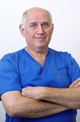
ד"ר גרי כהן
מנהל המרפאה
בוגר אוניברסיטת תל אביב מחזור 1990.
חבר פעיל בארגונים להשתלות דנטליות בארצות הברית ואירופה. ביצע עשרות אלפי שתלים ושיקום על גבי שתלים משנת 1990 ועד היום.
מוסמך לטפל בהרדמה כללית.
מרצה, כותב מאמרים ושותף בצוות בינלאומי לפיתוח השתלות דנטליות.
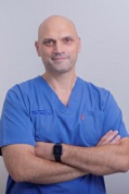
ד"ר אנג'י מאיר
רופא שיניים
בוגר האוניברסיטה העברית משנת 2002. עוסק בכל תחומי רפואת השיניים עם דגש מיוחד לתחום השיקום ואסתטיקה דנטלית.
משתלם במרכז לאסתטיקה בהדסה.
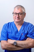
ד"ר בריס דני
רופא שיניים
בוגר האוניברסיטה העברית הדסה 1987. בעל ניסיון רב בשיקומים מורכבים ושיקום על שתלים, בהדגשת האדם כמרכז הטיפול. מורשה מטעם משרד הבריאות לטיפולי שיניים בהרדמה כללית. דובר עברית, אנגלית וספרדית, אבל מקשיב בכל השפות.
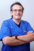
ד"ר פרנק אלוש
רופא שיניים
בוגר אוניברסיטת פריז 5 -1996. עוסק ברפואת שיניים כללית עם דגש לשיקום הפה.
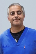
ד"ר רמי טביב
מומחה בכירורגיה פה ולסת
בוגר האוניברסיטה העברית הדסה עין כרם. מומחה בכירורגית פה ולסתות ורופא בכיר באוניברסיטה העברית ובית החולים הדסה עין כרם.

ד'ר שמואל טאו
D.M.D ירושלים 1974 הפקולטה לרפואת שיניים.
M.dent(witz) 1983 אוניברסיטת וויטוואטרסראנד יוהנסבורג דרום אפריקה.
1982 מומחה בשיקום הפה מטעם המועצה המדעית ישראל.לימד בפקולטה לרפואת שיניים בירושלים וכעת מלמד בבית הספר על שם גולדשלאגר אוניברסיטת תל אביב.
1988-2016 יועץ ארצי לשיקום הפה ברשת מירפאות מכבי דנט.
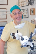
ד"ר טיירי לסקר
רופא שיניים
רופא שיניים לטיפולי שורש.
בוגר אוניברסיטת פריס 5 – 1995.
מדריך באוניברסיטת Garancière צרפת במחלקה לטיפולי שורש 1998-2012.
חבר באיגוד הצרפתי לטיפולי שורש ( SFE).

אולגה אבייב
רופאת שיניים
בוגרת אוניברסיטת וורונז' מדיקל ברוסיה - 2009.
עוסקת ברפואת שיניים כללית עם דגש על כירורגיה,
משמרת ושתלים.

ד"ר גיא טרטקובסקי
אנדודונט
סיים את לימודי רפואת השיניים בשנת 2009 בהדסה עין כרם. סיים התמחות באנדודונטיה (טיפולי שורש) בשנת 2019. חבר בהר"ש.
חבר באיגוד הישראלי לאנדודנטיה חבר באיגוד האמריקאי לאנדודונטיה AAE
דובר עברית, רוסית, אנגלית ברמת שפת אם ומעט צרפתית.

ד"ר טל בסיאלוב
רופא שיניים
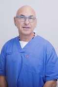
ד"ר דורון קרן
רופא שיניים
בוגר אוניברסיטת תל-אביב מחזור 1991. עוסק ברפואת שיניים כללית עם דגש בשיקום.

ד"ר נתלי פרומקין
רופאת שיניים, מומחית לפריודונטיה
בוגרת האוניברסיטה העברית הדסה עין כרם 2006. במקביל קיבלה את התואר המוסמך במדעי הטבע בחוג לגנטיקה של האדם. מומחית במחלות חניכיים התמחתה בפקולטה לרפואת שיניים של האוניברסיטה העברית והדסה.
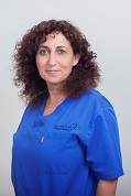
ד"ר יעל הרוש
רופאת שיניים
בוגרת אוניברסיטת בריסל 1992. עוסקת ברפואת שיניים כללית, שיקום ואסתטיקה

ד"ר קורין בואזיז
רופאת שיניים
בוגרת אוניברסיטת
Louis Pasteur de Strasbourg שנת 1991.
עוסקת בכל תחומי רפואת השיניים עם דגש לטיפולי שיניים למטופלים הסובלים מחרדה דנטלית גבוהה וילדים וגישה הוליסטית.

ד"ר איתי שימרון
רופא שיניים
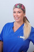
ד"ר יסניה קצנטיני
רפואת שיניים
1992-2001 לימודי רפואת שיניים באוניברסיטה בפרו
2009 עלתה לארץ ומאז עוסקת ברפואת שיניים כללית עם דגש על כרורגיה ושיקום הפה.
עוסקת גם באסתטיקה (הזרקות בוטוקס וחומצה אילרואית)
דוברת עברית וספרדית
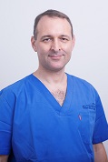
ד"ר אור אופק
אורתודונט
מומחה ליישור שיניים ולסתות - הדסה עין כרם.
בוגר הפקולטה לרפואת שיניים באוניברסיטה העברית 2006 (DMD).
בעל דוקטוראט מחקרי (PhD) בביולוגיה של העצם 2009.
מתמחה במגוון שיטות מתקדמות ומתוחכמות: גלויות ו׳בלתי נראות׳ כגון סמכים ננעלים, סמכים שקופים או פנימיים ופלטות שקופות.
מדריך מתמחים במחלקה בהדסה עין כרם. חבר באגודה האורתודונטית הישראלית ובהסתדרות הרפואית.
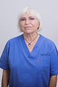
ד"ר מירה מרקוביץ
רופאת שיניים

ד"ר סהר נדל
רופאת שיניים מומחית בכירורגית פה ולסת
צוות שינניות במרפאה של ד"ר ג'רי כהן

דנה רז
שיננית מוסמכת
שיננית מוסמכת , הדסה, ירושלים 1999.
בעלת תואר ראשון בפסיכולוגיה.
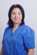
נילי גואל
שיננית מוסמכת
שיננית מוסמכת, מכללת רמת גן 2005.
1992 – קורס סייעות.

ז'אנה קנטרוביץ
שיננית מוסמכת
בוגרת מכללת רמת גן 2017
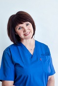
אירינה מגילבסקי
שיננית מוסמכת
שיננית מוסמכת, אוניברסיטת ת"א 2011.
2002 - קורס סייעות.
צוות המרפאה

ישראל גרשון
מנהל המרפאה
ניהל מרפאות ציבוריות בתחום רפואת השיניים משנת 1988.

ליאת
מנהלת שירות לקוחות
רפלקסולוגית

חיה שקד
רפלקסולוגית
רפלקסולוגית, נטורופטית מוסמכת N.D,
הרבליסטית קלינית CL.H
רפלקסולוגית בכירה
בוגרת מכללת סמינר הקיבוצים, סיימה בהצטיינות.
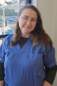
אירנה
רפלקסולוגית בכירה
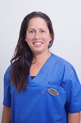
שרון אלימלך
רפלקסולוגית בכירה
סייעות
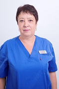
אנה שטרקמן
סייעת אחראית
2003 קורס סייעות ומזכירות רפואית.

אורטל דברייב
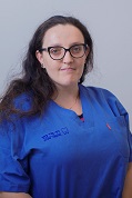
אלכסנדרה שכטר

אנה קנטרוביץ
אחראית תחום כירורגיה
2004 קורס סייעות לרופא שיניים

לודמילה קפוסטין

אנה אולשנסקי
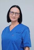
יוסיפוב מרינה
אחראית תחום שתלים ואחראית קומה שנייה
2005 קורס סייעות ומזכירה רפואית
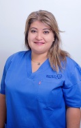
מורן פרהן
מ"מ אחראית קומה ראשונה
סייעת מוסמכת בעלת ותק של 15 שנה

סמדר שלמה
מזכירות רפואיות
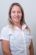
ילנה איברגימוב
אחראית לו"ז רופאים
מזכירה רפואית במרפאת שיניים
משנת 2000
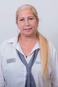
כהן מירי
אחראית מעקב מטופלים
מזכירה רפואית במרפאת שיניים משנת 2000.

איריס וטורי
מזכירה רפואית
במרפאת שיניים משנת 1992.

שלומית יצחקי

אשר תמי
אחראית מעקב מטופלים
2004 - קורס מזכירות רפואית

עדנה בוצר
אחראית מעקב מטופלים
מזכירה רפואית במרפאת שיניים משנת 1990 .
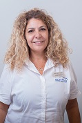
אסתר ינקו סגלוביץ'
מזכירה רפואית, אחראית אורתודונטיה
במרפאת שיניים מ- 2006
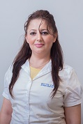
אנג'לה יצחקוב

דניאל סיטון

